All published articles of this journal are available on ScienceDirect.

Emerging Neurobiomarkers in Neurodegenerative Disorders: From Molecular Mechanisms to Clinical Translation
Abstract
Neurodegenerative disorders such as Alzheimer’s Disease (AD) and Parkinson’s Disease (PD) are characterized by a progressive loss of neurons, correlating with loss of motor and cognitive function that has an impact on global health. Diagnosis of these diseases in clinical settings is generally delayed due to reliance on clinical features and diagnostic criteria that focus on symptomatic stages of the neurodegenerative disorder. Growing evidence suggests that pathological activity at a molecular level can occur years before the onset of symptoms. This narrative review focuses on recent progress in fluid, imaging, genetic, and multi-omics as well as digital biomarkers, with an emphasis on their diagnostic implementation, feasibility, value, and translation into clinical settings. In AD, cerebrospinal fluid and plasma biomarkers such as Glial Fibrillary Acid Protein (GFAP), Aβ1–42/Aβ1–40 ratios, and phosphorylated tau isomers (p-tau181, p-tau217) have been shown to have promising diagnostic relevance and utility. Protein Misfolding Cyclic Amplification (PMCA) and Real-Time Quaking-Induced Conversion (RT-QuIC) have shown high diagnostic specificity for detecting α-synuclein seeding activity, accurately distinguishing synucleinopathies, which are of particular interest in PD. Corresponding imaging biomarkers, including structural and functional MRI, Positron Emission Tomography (PET), and multimodal imaging, provide complementary insight into the relevance of disease-stage categorization. Multi-omics profiling and digital biomarkers, including wearable sensors, speech and gait analysis, and electroencephalography combined with artificial intelligence-driven pattern recognition, further broaden the biomarker terrain toward continuous, precision-based monitoring of neurodegeneration. Despite significant progress, limitations remain in standardization, biological heterogeneity, and validation of relevant findings. Ethical considerations related to preclinical diagnosis also require considerable premeditation. The change toward the implementation of a multimodal framework has the potential to shift neurodegenerative disease diagnosis from symptomatic late-stage diagnosis to early, molecularly mechanistic-driven medical aftermath.
1. INTRODUCTION
Neurological diseases have been found to have an impact on global health, remaining a leading cause of death worldwide. There has been a decrease in age-standardized rates; however, the total number of deaths has increased across the years of 1990-2016. PD, Multiple Sclerosis (MS), and other motor neuron diseases have been shown to follow increasing prevalence rates between world regions. Key information also indicates that socioeconomic development does not aid in the determination of the extent of prevalence of AD [1]. Increasing global rates in cases of PD incidence, mortality, and prevalence have been observed in individuals aged 55 and older from 1991 to 2021. Taking age-stratified analyses into account, there has been a significant increase in PD burden amongst the elderly, aged 95 years and older, highlighting aging as the fundamental risk factor for neurodegenerative diseases. Prevalence and incidence were found to be at an all-time high across East Asia. Deaths across the Asia Pacific region, which has a high income margin, showed the greatest increase in annual deaths [2]. Individuals with PD and their respective caregivers underwent a significant and increasing economic burden as their disease progressed, with an increased use of healthcare resources in the US and Europe as compared to Japan. The financial constraint shown leads to an emphasis on comprehensive disease control at the early stages. People with an advanced state of PD would have a greater cost that would affect them directly, as compared to those with an early state of the disease. A specialized team of healthcare personnel also needs to be provided for patients with such advanced stages of the disease. There is a rise in mean annual costs in patients with advanced stages of PD in comparison to those with an intermediate stage of the disease in the USA and Europe, along with a slight increase in Japan. Also, in individuals with an advanced PD stage, the main driving factors for high annual medical costs were found to be hospitalizations and medications that were prescribed [3].
Substantial diagnostic delays have been reportedly observed for patients with typical young-onset AD of 5.5 years. Despite accounting for age as the main confounding factor, delayed diagnosis is prevalent. Prolonged periods were also observed to be a significant factor in the duration between symptom onset and initiation of medical services, with an average of about 3.4 years. Physicians were supplied with MRI scans on the first visit, which were generally normal [4]. Diagnostic accuracy for Parkinson’s disease at the first specialist visit remains limited, with clinical diagnosis often unreliable in the early phase and increasingly accurate only after several years of symptom evolution.
A growing body of evidence shows that neurodegenerative pathological processes are active for a prolonged period of time before clinical diagnosis and onset of symptoms, producing an extension of the time of preclinical and prodromal disease. Plasma biomarker studies have shown that biomarkers such as NfL and activation of GFAP have been detected in healthy subjects before a neurodegenerative clinical diagnosis is made. Population-based data from the UK Biobank indicate that elevated baseline NfL is connected to an increased risk of diagnosis following it in PD, atypical Parkinsonian disorders, AD, and Amyotrophic Lateral Sclerosis (ALS) [5]. Plasma biomarkers such as phosphorylated tau-81 and GFAP have been associated with markers of future brain atrophy and cognitive change among unimpaired elderly adults and are being used to predict future neurodegenerative and cognitive pathways. Changes in cognitive function within Isolated/Idiopathic Rapid Eye Movements (iRBD) have been reported to commence up to 10 years before conversion to Dementia with Lewy bodies (DLB), beginning with a decrease in executive function along with attention, memory, and finally visuospatial dysfunction close to the time of conversion [6]. Associations were observed between baseline serum NfL levels at diagnosis and changes in NfL levels over 5 years of disease duration, with declines in cognitive and motor function in individuals with PD. This further supports the observation that changes in NfL levels may begin at the prodromal phase in PD [7]. Altogether, findings from cohorts measuring plasma biomarkers can be synthesized into a model reinforcing that changes in neurodegenerative disorders can initiate at the prodromal and preclinical stages before evolving into clinical diagnoses under current diagnostic criteria.
Neurobiomarkers have been gaining advancements in their discovery, showing significant correlation with neurodegenerative diseases such as PD, AD, ALS, Multiple Sclerosis, etc. Clinical transition remains fractured across domains such as molecular, imaging, genetic, and digital. Reviews tend to focus on disease-specific applicability, such as PD and AD; however, few synthesize the capacity to integrate a broad spectrum of insights into a translation to clinical settings across neurodegenerative diseases. With the impending rise in multi-omics, blood-based assays, and digital phenotyping tools, a framework that links neuropathological mechanisms to clinically translatable biomarkers is warranted.
This narrative review focuses on the ability to correlate emerging biomarkers with concurrently employed pathophysiological mechanisms and translate them into clinical diagnostic settings. It also evaluates the clinical competence of neurodegenerative biomarkers such as fluid, imaging, genetic, and digital biomarkers at an overall level rather than in isolation. It also determines and evaluates the challenges to be addressed for implementation, including factors such as standardization, validation, regulation, and global accessibility of these neurobiomarkers. This review takes a mechanistic and translational approach, with an emphasis on pathological mechanisms such as protein aggregation, neuroinflammation, synaptic dysfunction, and axonal injury, to assess their correlation with cerebrospinal fluid, blood, imaging modalities, multi-omics, and digital technologies. Rather than providing exhaustive information on disease-specific indices, this review focuses on providing insight to enhance precision neurology and interventional strategies in early disease states. By issuing relevant information, this review seeks to provide reasons for transitioning from symptom-based neurodegenerative syndromes to molecularly staged conditions, ultimately to support early diagnosis and therapeutic approaches to stop the disease at an early stage. This review proposes that various biomarkers such as molecular, fluid, imaging, multi-omics, and digital biomarkers can converge to form a staged, multi-diagnostic framework that may shift the neurodegenerative disease management from a reactive, symptom-based diagnostic protocol to early detection through informed molecular-based diagnosis, clinical trial enrichment, and precision monitoring.
1.1. Search Strategy
Databases such as PubMed, Scopus, Web of Science, and Google Scholar were searched using keywords like “neurodegenerative biomarkers,” “Alzheimer's fluid biomarkers,” “Parkinson's imaging biomarkers,” “neurofilament light chain,” “phosphorylated tau,” “digital biomarkers neurodegeneration,” and “multi-omics neurodegeneration” since January 1995 through January 2026, including peer-reviewed original research, systematic reviews, meta-analyses, and key clinical guidelines limited to the English language. Studies were selected based on relevance, recency, and contribution to the translational theme rather than through PRISMA-based screening. Conference abstracts without full publications, editorials without primary data, and non-English publications were excluded from the search.
1.2. Biomarker Readiness Classification
To consistently evaluate the translational maturity of the biomarkers in this review, each biomarker is classified according to a five-tier readiness scheme, including stages such as discovery, analytical validation, clinical validation, regulatory validation, and routine clinical use. Firstly, the discovery stage includes biomarkers identified in exploratory or preclinical studies, with limited or no validation in human cohorts. Secondly, the analytical validation stage encompasses biomarkers that have demonstrated assay reliability, reproducibility, and technical performance, but the diagnostic value of these biomarkers is not confirmed in independent cohorts. Thirdly, the clinical validation stage includes biomarkers that have demonstrated diagnostic or prognostic performance across independent, adequately powered clinical cohorts. Fourthly, the regulatory qualification stage includes biomarkers that have formally gained approval by regulatory agencies such as the Food and Drug Administration for use in clinical trial enrollment or as diagnostic aids. Lastly, the routine clinical use stage includes biomarkers that have been incorporated into standard clinical practice guidelines with established reimbursement and accessibility pathways.
2. MOLECULAR BASIS OF NEURODEGENERATION: THE ORIGIN OF BIOMARKERS
2.1. Protein Misfolding and Aggregation (Tau, α-Synuclein, TDP-43)
A PET imaging study in healthy elderly individuals explored the uptake patterns of an in vivo biomarker for the accumulation of phosphorylated tau. Consistent anatomical patterns were observed with tau accumulation starting in the Medial Temporal Lobe (MTL), which would be correlated with specific Braak-like staging progressions that were measured using a computational method correlating closely with changes in cognitive measures [8]. Phosphorylated tau isoforms, particularly p-tau181 and p-tau217, accumulate as a downstream consequence of amyloid-driven tangle formation and are among the most extensively characterized fluid biomarkers of this pathway [9, 10]. Their diagnostic performance as CSF and plasma biomarkers is discussed in detail in Section 3.1 and Section 3.2. In patients with iRBD, CSF α-synuclein seeding assays demonstrated that individuals who tested positive had a higher correlation with the risk of developing PD or DLB than those who tested negative. This demonstration predicted, up to almost a decade before the clinical diagnosis of PD and DLB, in the reported individuals [11, 12]. These assays demonstrate the ability of CSF α-synuclein seeding assays to be translationally used as a biomarker in clinical settings. In studies concerning ALS and frontotemporal dementia, correlations between plasma TDP-43 and plasma in the disease group underscore an underlying pathology among these neurodegenerative diseases that requires further exploration [13, 14]. Among these biomarkers, plasma p-tau181, p-tau217, and CSF α-synuclein seeding assays have reached the clinical validation stage, while plasma TDP-43 remains at the discovery stage pending confirmation from larger, independent cohorts [9-14].
Overall, these findings underscore the importance of evaluating misfolded protein species not only as hallmarks of these disease states but also as disease-stage biomarkers.
2.2. Neuroinflammation and Glial Activation
Astrocytic reactivity in response to amyloid pathology is captured by GFAP, released into both CSF and plasma as a marker of glial activation [15]. GFAP's diagnostic performance across the AD continuum is discussed in Section 3.1 and Section 3.2. Microglial activation, as observed using translocator protein PET imaging, demonstrated increased binding in preclinical early AD [16]. This supports the implication of microglial activation being an early marker of AD. Inflammatory cytokines and chemokines in CSF and plasma are associated with neurodegenerative injury. Several inflammatory mediators, including interleukins and chemokines, have been described as elevated in CSF and plasma in association with neurodegenerative pathology, supporting a link between neuroinflammation and disease progression. Plasma and CSF GFAP have reached the clinical validation stage as early markers of astrocyte reactivity, whereas microglial activation, assessed by translocator protein PET imaging, and CSF cytokines remain at the discovery stage [15-18]. Neurodegeneration can be detected through neuroinflammation, an early biomarker that is demonstrated collectively through these findings.
2.3. Synaptic Dysfunction and Axonal Injury
Synaptic loss is observed to correlate with impairment across neurodegenerative disorders. Synaptic biomarkers such as neurogranin have been described as elevated in the CSF of patients with Alzheimer’s disease, supporting the evaluation of synaptic loss as a marker of neurodegeneration. Axonal injury releases Neurofilament Light Chain (NfL) into CSF and blood, and it is now one of the most widely measured markers of neurodegeneration across disease categories. Its diagnostic accuracy and, critically, its limitations for differential diagnosis are discussed in detail in Section 3.1 and Section 3.2.
In neurodegenerative disorders such as Huntington’s disease, elevated NfL concentrations have been reported years before clinical onset and correlate with subsequent motor and cognitive decline, supporting the notion that axonal injury is measurable during preclinical stages [19]. NfL has reached the clinical validation stage as a marker of axonal injury in PD. Collectively, these findings support NfL as a neurobiomarker of axonal injury that correlates with neurodegeneration in disease states, warranting its evaluation in clinical settings.
2.4. Mitochondrial Dysfunction and Oxidative Stress
Mitochondrial dysfunction is a fundamental cause of neurodegeneration by compromising cellular energy attained through metabolism and producing reactive oxygen species. In individuals with AD and progressive supranuclear palsy, metabolic analyses found oxidative stress that was associated with lipid metabolism and mitochondrial dysfunction, which is connected to lysine metabolism. This demonstrates that disruption of mitochondrial and metabolic pathways is a hallmark of neurodegeneration [20].
In AD, metabolites involved in energy regulation, neurotransmission, and mitochondrial function are generally found to be affected. Significant changes were reported in alanine, aspartate, and glutamate metabolisms in most brain regions, which were linked to mitochondrial dysfunction, impaired energy metabolism, and changes in mitochondrial function. This shows direct and indirect connections to neurodegeneration in AD, which warrants evaluation for consideration as potential clinical biomarkers [21].
Oxidative stress-related metabolites and CSF-based inflammatory markers were associated with biomarkers of general AD pathology [22]. This finding demonstrates that oxidative stress metabolites are related to neurodegeneration in AD. Oxidative stress biomarkers in individuals with Mild Cognitive Impairment (MCI) were also shown to be associated with neurodegeneration, ultimately leading to cognitive decline. Although a personalized approach may be warranted due to significant changes in age and sex that influence oxidative stress levels. Biomarkers for mitochondrial dysfunction and oxidative stress are currently at the discovery stage [20-22].
These findings collectively demonstrate that mitochondrial dysfunction and oxidative stress, both due to associated metabolites, caused neurodegeneration through direct and indirect processes, which underscores the need for their clinical evaluation.
2.5. Genetic and Epigenetic Influences
Genetic mutations are correlated with disease onset and are also associated with related neurodegenerative biomarker outcomes. In Huntington’s disease mutation carriers, plasma NfL concentrations have been described as elevated well before the emergence of clinical symptoms, supporting its potential as a preclinical prognostic marker [19]. Elevations in biomarkers preclinically were also reported similarly in a familial ALS cohort analysis [23].
Large-scale epigenomic analysis demonstrates that methylation-related cfDNA biomarkers can distinguish individuals with ALS from healthy individuals with high sensitivity. cfDNA biomarkers may also be able to predict ALS, but further studies are warranted to confirm this [24].
Interactions between genetic and epigenetic factors may also influence the onset and progression of disease. A genomic analysis identified specific CpG-related mutations that modify the age of onset in individuals with ALS [25]. This demonstrates wide implications, as epigenetic influences may play significant roles in the control of neurodegenerative diseases such as ALS.
Exploring beyond disease-specific mutations, changes in DNA methylation, SIRT activity, and BDNF expression are observed to decline significantly in patients with dementia or PD [26]. Their utility as potential epibiomarkers in neurodegenerative disorders is warranted, and further studies are required to determine their efficacy in the diagnosis of neurodegenerative disorders. Most genetic and epigenetic biomarkers, such as cfDNA and DNA methylation, are in the discovery stage and require further study [23, 25, 26].
Overall, these findings collectively demonstrate that considerations of genetic mutations and epigenomics need to be evaluated clinically to serve as effective biomarkers for diagnosing neurodegenerative diseases.
Protein misfolding, neuroinflammation, synaptic dysfunction, axonal injury, and mitochondrial dysfunction are interlinked mechanisms that do not occur in isolation but represent converging pathways that drive neurodegeneration from pre-clinical early stages of these diseases. Each mechanism provides distinct but measurable biomarkers. Misfolded proteins and inflammatory mediators are detectable in CSF and blood; axonal injury is assessed via NfL; metabolic dysfunction and oxidative stress are interlinked mechanisms; and epigenetic disturbances are increasingly accessible via omics platforms.
Pathophysiological Pathways and Corresponding Biomarker Sources are indicated in Fig. (1).
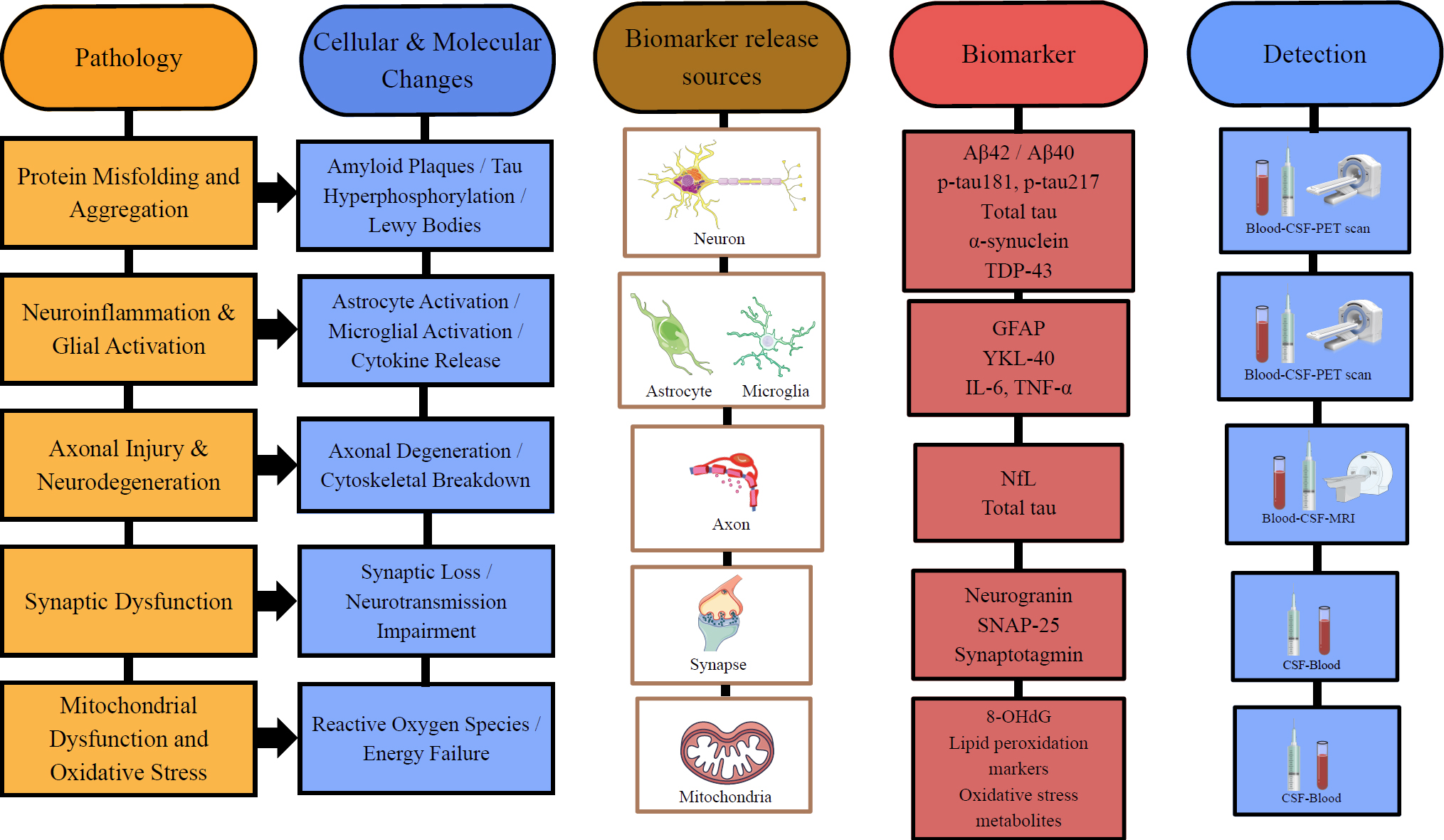
Pathophysiological pathways and corresponding biomarker sources.
3. FLUID BIOMARKERS: ADVANCING MINIMALLY INVASIVE DIAGNOSIS
3.1. Cerebrospinal Fluid Biomarkers
CSF biomarkers are among the most established indicators of neurodegeneration because they directly reflect changes in the central nervous system. They provide evidence of degenerative changes, including protein aggregation, neuronal injury, axonal dysfunction, and neuroinflammation, thereby promoting early detection of neurodegenerative disorders such as Alzheimer’s Disease, Parkinson’s Disease, Amyotrophic Lateral Sclerosis, and Frontotemporal Dementia (FTD). A comparative summary of the CSF, blood-based, and peripheral fluid biomarkers discussed throughout this section is presented in Table 1.
| Biomarker | Disease Context | Sample Type | Biological Mechanism | Clinical Application | Validation Stage | Major Limitations | Key References |
|---|---|---|---|---|---|---|---|
| Aβ42/Aβ40 ratio | AD | CSF, plasma | β-amyloid plaque deposition; reduced Aβ42 reflects cerebral sequestration into plaques | Amyloid-status confirmation; patient selection for anti-amyloid therapy | Clinical Validation | Inter-laboratory assay variability; lack of universally harmonized cutoffs; plasma ratio narrower than CSF | [27] |
| Total tau (t-tau) | AD, other tauopathies | CSF | Marker of general neuronal/axonal injury, not tau-specific pathology | Supportive marker of neurodegeneration severity | Clinical Validation | Non-specific; elevated in acute injury and multiple unrelated conditions | [28] |
| p-tau181 (CSF) | AD | CSF | Tau phosphorylation at threonine-181; reflects early tangle formation | Diagnostic aid; correlates with amyloid pathology | Clinical Validation | Later-stage marker relative to p-tau217; moderate specificity for AD vs. other dementias | [9] |
| p-tau181 (plasma) | AD | Plasma | Same mechanism as CSF p-tau181, peripherally sampled | Minimally invasive AD screening | Clinical Validation | Peripheral pre-analytical variability (fasting, storage, time-of-day); lower diagnostic accuracy than p-tau217 | [9] |
| p-tau217 (CSF) | AD | CSF | Tau phosphorylation at threonine-217; strongly correlates with tau PET/amyloid PET | High-accuracy diagnostic and staging marker | Clinical Validation | Requires lumbar puncture; assay standardization still evolving across platforms | [10] |
| p-tau217/Aβ42 ratio (plasma) | AD | Plasma | Combined amyloid/tau phosphorylation signal | Blood-based aid for amyloid-plaque detection in symptomatic adults | Clinical Validation | Not a stand-alone diagnostic; indeterminate-result rate; validated primarily in symptomatic, age-restricted cohorts; multi-lab reproducibility still consolidating | [10, 73] |
| α-synuclein SAA/RT-QuIC | PD, DLB, MSA | CSF | Detects seeding/aggregation of pathological α-synuclein via conformational amplification | Distinguishes synucleinopathies from healthy controls and other parkinsonism | Clinical Validation | Invasive sampling; assay still largely confined to specialized/research laboratories; imperfect cross-subtype specificity | [12, 31, 34, 35] |
| NfL (CSF) | AD, FTD, ALS, PD, MS, HD | CSF | Marker of axonal cytoskeletal injury | Disease progression and severity monitoring | Clinical Validation | Disease-nonspecific; elevated in any neuroaxonal injury, including non-neurodegenerative causes | [8, 29] |
| NfL (plasma) | AD, FTD, ALS, PD, MS, HD | Plasma | Same mechanism, peripherally sampled | Longitudinal monitoring; used as supportive (non-qualified) evidence in one ALS accelerated drug approval | Clinical Validation | Not formally biomarker-qualified (ref 81); lacks disease specificity; influenced by age and renal function | [8, 72] |
| GFAP (CSF/plasma) | AD | CSF, plasma | Intermediate filament protein reflecting reactive astrogliosis | Early marker of amyloid-related glial activation | Clinical Validation | Elevated non-specifically in other neuroinflammatory and injury states; cross-platform standardization incomplete | [5, 15] |
| YKL-40 | AD | CSF | Glycoprotein expressed by reactive astrocytes/microglia | Supportive neuroinflammatory marker | Analytical Validation | Single-study elevation data; no established diagnostic cutoff | [32] |
| TDP-43 / pTDP-43 (plasma/CSF) | ALS, FTD | Plasma, CSF | Cytoplasmic mislocalization/aggregation of TDP-43 proteinopathy | Investigational marker of underlying ALS/FTD pathology | Discovery | Weak plasma-CNS correlation; assay reproducibility not yet established | [13, 14] |
| Exosomal p-tau/amyloid proteins | AD | Plasma (neuron-derived exosomes) | Enrichment of pathological proteins in neuron-derived extracellular vesicles | Investigational early-detection marker | Discovery | Single-cohort evidence; complex, non-standardized isolation methods | [35] |
| Salivary α-synuclein | PD | Saliva | Reflects peripheral/CNS α-synuclein pathology via minor salivary glands | Investigational non-invasive screening tool | Analytical Validation | Single pilot cohort; unclear correlation with CNS disease burden | [36] |
| Urinary metabolites (8-OHdG) / cytokines | PD, AD | Urine | Oxidative DNA damage and peripheral inflammatory signaling | Investigational non-invasive monitoring | Discovery | Pilot-study evidence only; no standardized collection protocol; requires RCT-level confirmation | [37, 38] |
| Peripheral inflammatory mediators (IL-6, IL-1β, TNF-α, CRP) | AD, PD | Serum, plasma | Systemic inflammatory response accompanying neurodegeneration | Adjunct marker of neuroinflammatory burden | Analytical Validation | Highly non-specific; confounded by comorbid systemic inflammation | [39-42] |
3.1.1. Aβ42/Aβ40 Ratio
Amyloid-beta (Aβ) peptides are substances derived from the Amyloid-beta Precursor Protein (APP), which is cleaved by beta and gamma secretases to form Aβ. Amyloid-beta proteins, in particular Aβ42 and Aβ40, are the main biomarkers that are observed to cause pathologic abnormalities, generally in AD. They deposit as plaques in CSF, with Aβ40 being relatively stable. Concentrations of CSF Aβ42 are reduced in Alzheimer’s disease as the peptide is sequestered into cerebral amyloid plaques, and lower CSF Aβ42 concentrations correspond to greater brain amyloid deposition. Aβ42 concentration levels may be influenced by individual variability in amyloid production, and therefore the Aβ42/Aβ40 ratio serves to standardize Aβ42 levels to total amyloid production levels, improving diagnostic capability. A study conducted by Lewczuk et al. (2014) demonstrated that the CSF Aβ42/Aβ40 ratio is significantly better than the isolated use of Aβ42 concentration levels for diagnostic accuracy [27]. CSF Aβ42/40 ratio is currently at the clinical validation stage [27]. Amyloid-beta peptides are well-established as robust biomarkers for detecting early neurodegenerative disease.
3.1.2. Total Tau and Phosphorylated Tau
Total tau (t-tau) and phosphorylated tau (p-tau) are CSF biomarkers that correlate with neuronal injury and neurologic pathology. T-tau is released into the CSF after damage to neurons and axons, and its release is associated with the intensity of neurodegeneration. CSF t-tau is elevated from the earliest clinical stages of Alzheimer’s disease, supports diagnostic discrimination between AD and cognitively unimpaired individuals, and is associated with concurrent amyloid pathology and longitudinal decline in cognition and brain structure. CSF p-tau217 shows even greater disease specificity: levels increase approximately 6-fold in AD patients compared with only 1.3-fold for p-tau181, and CSF p-tau217 identifies amyloid-positive individuals with sensitivity and specificity both exceeding 90% (AUC 0.961), indicating its capability to be used adjunctively in the diagnosis of AD [28]. Elevated CSF t-tau and p-tau levels at baseline are also associated with a substantially increased risk of subsequent clinical decline and conversion to AD dementia, underscoring their use in the prognosis of AD. CSF t-tau, p-tau181, and p-tau217 are currently at the clinical validation stage [9, 10].
3.1.3. Neurofilament Light Chain (NfL)
Neurofilament Light Chain (NfL) is an intermediate filament protein that is concentrated at neuronal axons and is released into the CSF following axonal injury. Elevated levels of CSF NfL correlate with direct axonal injury and neurodegeneration. NfL has been shown to be released into the CSF after axonal damage, establishing its role as a biomarker for neuronal injury, and has reached the clinical validation stage, as mentioned previously. Neurodegenerative disorders have elevated CSF NfL concentrations in FTD, correlating with progression and severity of the disease. Scherling et al. (2014) reported elevated CSF NfL concentrations in patients with FTD, showing associations with disease progression and severity of disease state [29]. Increased levels of NfL generally occur in the early stages of the disease. NfL concentrations were reportedly increased years before the onset of symptoms, suggesting that axonal injury begins at preclinical phases of neurodegeneration [30].
3.1.4. α-Synuclein
α-Synuclein is a presynaptic protein that plays an important role in synucleinopathies such as Parkinson’s disease and dementia with Lewy bodies. It is abundant in the brain, primarily regionalized to the axon terminals of presynaptic neurons. Aggregation of α-synuclein leads to neuronal dysfunction in PD and DLB, eventually causing neurodegeneration. Across independent cohorts, CSF α-synuclein Seed-amplification Assay (SAA) has demonstrated sensitivity of 90–97% and specificity of 87–100% for PD and DLB, with comparable performance (90% sensitivity, 90% specificity) in prodromal iRBD cohorts years before clinical diagnosis [12, 31, 32].
3.1.5. Glial and Inflammatory Markers
Glial activation, as reflected by GFAP, and neuroinflammation, which is expressed through inflammatory markers such as cytokines and chemokines, are important factors that cause neurodegenerative disease progression. CSF biomarkers that correlate with such processes are reflective of disease stages. GFAP is an intermediate filament protein that is expressed in astrocytes during the early reactive state of disease, serving as a potential biomarker of the preclinical disease state. CSF GFAP has been reported to exhibit more modest discriminative performance for amyloid positivity across the Alzheimer’s disease continuum than its plasma counterpart [16]. YKL-40, a glycoprotein expressed by microglia and astrocytes in an activated state, is reportedly increased in neurodegenerative diseases and may be associated with inflammation in the central nervous system. CSF YKL-40 concentrations have been reported to be increased in Alzheimer’s disease and associated with glial activation, but remain at the analytical validation stage and require further standardization [17].
Despite their diagnostic value, CSF-based fluid biomarkers have several limitations, including the invasiveness of lumbar puncture, limiting repeat sampling, and inter-laboratory assay variability. These limitations prevent universal diagnostic cutoffs for markers such as Aβ42/40, p-tau181/217, and NfL [27, 28]. NfL is additionally confounded due to lack of disease specificity and is included in non-neurodegenerative conditions like stroke and active neuroinflammation [33].
3.2. Blood-Based Biomarkers
The diagnosis and monitoring of neurodegenerative disorders have had significant advancements lately, from the emerging studies of blood-based biomarkers. Blood-based biomarkers may substitute for CSF biomarkers and neuroimaging due to their minimal intracranial invasion.
Blood-based biomarkers are major advancements in the diagnosis and monitoring of neurodegenerative disorders. They are minimally invasive, reliable alternatives to CSF and neuroimaging. Technological improvements in ultra-sensitive immunoassays have enabled reliable detection of less-abundant neuronal and glial proteins in plasma and serum. Blood-based biomarkers, therefore, reflect important pathological processes such as protein aggregation, axonal injury, and neuroinflammation.
3.2.1. Plasma Phosphorylated au soforms
Isoforms of plasma phosphorylated tau (p-tau) have shown strong disease specificity and are diagnostically applicable. They are particularly relevant to Alzheimer’s disease but also have broader utility in neurodegenerative disorders. Palmqvist et al. (2020) demonstrated that plasma p-tau217 levels increased in the early stage of the disease and accurately distinguished AD from other neurodegenerative disorders. Plasma p-tau217 discriminated AD dementia from other neurodegenerative diseases with an AUC of 0.96 (95% CI, 0.93–0.98; sensitivity, 93%; specificity, 88%), and the p-tau217/Aβ42 ratio performed comparably (AUC, 0.94; sensitivity, 82%; specificity, 92%). Plasma p-tau181 showed weaker discrimination (AUC 0.81; sensitivity 50%, specificity 89%), underperforming its CSF counterpart and plasma p-tau217 alike [10].
In an independent cohort, plasma p-tau181 alone discriminated AD dementia from non-AD neurodegenerative diseases with an AUC of 0.93 (sensitivity 92%, specificity 87%), performing similarly to CSF p-tau181 and tau-PET and outperforming CSF Aβ42/Aβ40 and CSF t-tau in the same cohort [9]. Plasma p-tau181 also predicted abnormal tau-PET status, with an AUC of 0.86 (sensitivity 80%, specificity 77%) for early-stage (Braak I–IV) tau pathology and 0.90 (sensitivity 93%, specificity 79%) for late-stage (Braak V–VI) tau pathology [9], supporting its potential use as a pre-screening tool to identify patients who would benefit from confirmatory PET or CSF testing.
Plasma p-tau181 is additionally one of the few blood-based AD biomarkers validated directly against neuropathology: in an autopsy-confirmed cohort, antemortem plasma p-tau181 accurately distinguished pathologically confirmed AD from non-AD cases (AUC 0.85), providing histopathological rather than purely PET- or CSF-based confirmation of its diagnostic validity [9].
Beyond cross-sectional diagnosis, plasma p-tau181 also predicted longitudinal progression to AD dementia in cognitively unimpaired and MCI individuals over up to 8 years of follow-up, with each standard-deviation increase in baseline plasma p-tau181 associated with a hazard ratio of 3.8 for future AD dementia, comparable to CSF p-tau181 (HR 3.9) [9]. When plasma p-tau181, T-tau, Aβ42/Aβ40, and NfL were entered together in a multivariable model, only p-tau181 remained independently associated with AD dementia risk; none of plasma T-tau, Aβ42/Aβ40, or NfL retained independent prognostic value [9].
Phosphorylated tau is therefore shown to be a reliable biomarker that has reached the clinical validation stage and reflects the concurrent disease state.
3.2.2. Plasma Neurofilament Light Chain (NfL)
Neurofilament Light Chain (NfL) is a component of the cytoskeleton of axons, emerging as a biomarker of neuronal injury across multiple neurodegenerative disorders. Plasma NfL concentrations are increased in AD, FTD, ALS, PD, and HD. Plasma NfL levels have been observed to be significantly higher in individuals with neurodegenerative disease compared to healthy individuals. Additionally, it correlated strongly with disease severity, brain atrophy, and progression of the disease state, reaching the clinical validation stage [34].
Plasma and CSF NfL levels show a strong correlation, suggesting that plasma measurements are highly associated with axonal injury of the central nervous system. Plasma NfL is thus shown to be a biomarker for neurodegeneration that warrants evaluation of its utility in disease monitoring and prodromal diagnosis in neurodegenerative disorders. Plasma NfL illustrates a critical distinction between detecting neurodegeneration and diagnosing a specific disease. In the BioFINDER-2 cohort, plasma NfL performed no better than chance when distinguishing AD dementia from other neurodegenerative diseases (AUC 0.50, 95% CI 0.42–0.58, not statistically significant; sensitivity 67%, specificity 38%) [10]. This lack of specificity is corroborated by an entirely different study design: in the multivariable Cox regression model described above for plasma p-tau181 [9], NfL was not independently associated with AD dementia risk, reinforcing that its elevation reflects the presence of neurodegeneration rather than any AD-specific process. Multicentre validation data further show that NfL's accuracy varies sharply depending on which conditions are being compared: it achieves only moderate accuracy separating AD dementia from cognitively unimpaired individuals (AUC 0.79–0.80; specificity 76–78% exceeding sensitivity 65–67%), performs considerably better distinguishing atypical parkinsonian disorders from PD (AUC 0.86–0.95; specificity 89–100%), but falls to an AUC of only 0.72 when distinguishing FTD from ALS, two conditions with similarly severe axonal injury [34]. At a cohort level, NfL identifies the presence of any neurodegenerative disorder against controls with good accuracy (AUC 0.83–0.90), but this accuracy drops substantially once PD patients are included in the neurodegenerative group (AUC 0.74–0.75), since PD itself produces comparatively modest NfL elevation [34]. Taken together, plasma NfL functions well as a general screening marker for neurodegeneration but should not be relied upon for differential diagnosis between specific conditions or for AD-specific risk prediction.
3.2.3. Plasma Glial Fibrillary Acidic Protein (GFAP)
Plasma Glial Fibrillary Acidic Protein (GFAP) is an intermediate filament protein that is expressed in astrocytes. Astrocyte activation and neuroinflammation are measurable by GFAP, as it serves as a biomarker. Plasma GFAP levels have been observed to be increased in AD and also correlate with amyloid proteins. Chatterjee et al. (2023) showed that plasma GFAP concentrations were significantly increased across the Alzheimer’s disease continuum in amyloid-positive individuals, including cognitively unimpaired individuals, indicating that astrocyte activation is detectable using plasma GFAP measurements from the earliest stages of the disease [15].
These observations highlight plasma GFAP as a potential biomarker that has reached the clinical validation stage and is relevant to neuroinflammation and linked to amyloid proteins [15].
3.2.4. Exosomal Biomarkers
Exosomes are a class of extracellular vesicles that serve as an additional source of disease-specific biomarkers. Exosomes contain proteins that originate from neurons and are associated with pathological processes of the brain. Fiandaca et al. (2015) demonstrated that plasma exosomes that were neuron-derived contained increased levels of p-tau and amyloid-related proteins in individuals who developed AD at a later stage. The biomarker changes were detected years before the onset of symptoms of AD, showing its potential utility as an early biomarker of the disease that reflects early pathology and presents as a biomarker at the discovery stage [35]. Exosomes offer a unique approach for detecting intracellular neurodegenerative processes in blood samples.
3.2.5. Ultra-sensitive Detection Platforms
Neurodegenerative low-abundance biomarkers improve with ultra-sensitive tests like Single Molecule Array (Simoa). These assays can detect proteins at significantly low concentration levels, resulting in better diagnostic accuracy. Ultra-sensitive immunoassays have been described as capable of reliably quantifying plasma phosphorylated tau, with strong correlations reported between plasma p-tau181, CSF, and imaging biomarkers, supporting its role as an emerging blood-based biomarker that requires further validation in larger analyses [11]. Blood-based biomarkers may become tools for diagnosis and clinical research through these ultrasensitive tests, which have made considerable technological progress.
Plasma biomarker levels can be influenced by hepatic or renal impairment, which may alter concentrations independently of neurodegenerative pathology. Standardized protocols for multiple pre-analytical factors such as fasting status, draw time, and sample handling that can affect measured biomarker levels are not yet uniformly applied.
3.3. Emerging Peripheral Biomarkers
Peripheral biomarkers are derived from sources such as saliva and urine, as well as other peripheral immune signatures. Detection of neurodegenerative disorders through peripheral biomarkers is gaining attention, as these biomarkers may be used in preclinical or early disease stages. Peripheral biomarkers are advantageous for their offers, such as minimal invasiveness, accessibility, and potential complementarity to CSF and plasma biomarkers. They may be used as a utility for insight into systemic and peripheral manifestations of neurodegenerative disorders.
3.3.1. Salivary and Urinary Biomarkers
Non-invasive biofluids such as saliva and urine can be collected repeatedly, making them ideal candidates for monitoring neurodegenerative disorders. These biofluids contain a wide range of biomarkers that are easily accessible. The collection of salivary samples does not require any specific skills from patients or medical staff. It is also not found to be dangerous to patients, making it collectively an alluring source of sample collection for biomarkers. Neuropathological and systemic processes can be examined in these biofluids, which contain a wide range of biomarkers, such as proteins and metabolites, that may reflect the underlying pathology of the disease.
Salivary biomarkers have been shown to be relevant to α-synuclein pathology. A study conducted by Vivacqua et al. (2019) demonstrated that total salivary α-synuclein was significantly lower in patients with PD than in healthy subjects. Salivary α-synuclein was also found to be able to differentiate between patients with Progressive Supranuclear Palsy (PSP) and patients with PD, where patients with PSP had higher α-synuclein levels, reportedly in comparison to patients with PD, supporting it as a biomarker at the analytical validation stage requiring further standardization [36]. This finding shows that salivary α-synuclein can be used clinically to differentiate patients with PSP from patients with PD.
Changes in urinary metabolites may signal early stages of PD. It is generally considered a favourable source of biomarkers, as it is non-invasive. Urinary 8-hydroxy-2’-deoxyguanosine (8-OHdG), a biomarker of DNA oxidative damage, has been shown to correlate with PD progression. Urine is a multifaceted biofluid that contains multiple metabolites that can be accessed as biomarkers for the prognosis and diagnosis of neurodegenerative disorders. It is also a more convenient sampling method than blood and CSF due to its accessible range. Although other body fluids have remarkable data, the rich content of urine from its metabolites, protein content, stability, and ease of access make it a valuable source for the exploration of biomarker discovery in neurodegenerative disorders [37]
Inflammatory mediators present in urine have shown potential for diagnostic capability in AD. A pilot study conducted by Saiyed et al. (2023) demonstrated significant changes in the concentrations of several urinary cytokines when subjects with AD were compared with healthy subjects. Mediators such as IL6ST and MMP-2 were identified as significantly changed in healthy subjects and subjects with AD. MMP-2 levels were observed to be elevated in AD as compared to healthy subjects. Expression of other inflammatory mediators was also identified, including TNFSF13, IL-2, IL-20, and IL-12, among others, which were observed in MCI compared with AD and healthy subjects [38]. These findings collectively identify urine as a potential source of biomarkers at the discovery stage, warranting investigation through randomized controlled trials to assess its diagnostic capability.
3.3.2. Peripheral Immune Signatures
Peripheral immune changes are an important source of biomarkers that are concurrently emerging in neurodegenerative disorders, representing inflammatory responses that correlate with their underlying pathology.
Elevated peripheral inflammatory mediators such as IL-6, IL-1β, TNF-α, and MCP-1 were found to be correlated with AD [39]. A large cohort examining newly diagnosed PD patients, conducted by Williams-Gray et al. (2016), demonstrated that elevated serum IL-6 levels may predict motor progression at an increased pace, indicating that selected immune biomarkers may reflect the severity of disease states and their progression [40].
The prediction of outcomes in neurodegenerative disorders may serve an important role in identifying stages of disease progression. Systemic inflammation was established to be a reproducible feature of PD, supporting that the peripheral immune activation response is correlated with neuroinflammation.
Multiple inflammatory mediators such as IL-6, IL-1β, TNF-α, and C-reactive Protein (CRP) were observed to be elevated in patients with PD compared to healthy controls [41]. These findings collectively establish the role of peripheral immune mediators and their importance to the diagnostic approach of neurodegenerative disorders.
Inflammatory markers are not mere phenomena that are used to assess the severity of diseases. A cohort study by Yazdani et al. (2019) concluded that elevations in leukocyte counts and systemic inflammatory markers were correlated with increased risk of developing PD [42]. Peripheral immune markers are the analytical validation stage requiring replication and standardization [39-42]. This finding suggests that dysregulation of peripheral immune signatures may precede the disease onset of neurodegenerative disorders.
Salivary and urinary biomarker studies remain limited to small, single-center cohorts with non-standardized collection protocols [36-38]. Their diagnostic relevance may be in question due to limited correlation with underlying CNS pathology.
A comparative summary of quantitative diagnostic performance metrics, including AUC, sensitivity, and specificity, for the key fluid biomarkers discussed in this section is presented in Table 2.
Comprehensively, the fluid biomarkers demonstrated substantially different degrees of translational maturity. CSF and plasma Aβ42/40, p-tau181, and p-tau217 show consistent progress in validation, with genuine cross-cohort performance supporting their use in diagnostic algorithms and clinical trial enrichment. NfL and GFAP serve as broad, informative biomarkers of axonal injury and glial activation, remain non-specific across disease states, and are better positioned for monitoring than as standalone diagnostic biomarkers. α-Synuclein seed-amplification assays are emerging as validated, disease-specific biomarkers. Peripheral salivary, urinary, and exosomal samples remain in the early stages and require analytical validation. Currently, fluid biomarkers present as strong candidates that, when paired with imaging biomarkers, can lead to a multimodal diagnostic framework that warrants further evaluation.
4. IMAGING BIOMARKERS
4.1. Structural MRI Patterns
MRI patterns offer hope in the early detection of neurodegenerative disorders and provide targeted treatment through the localization of brain structures. Generally, it is combined with other imaging biomarkers such as Computed Tomography (CT) and Positron Emission Tomography (PET) to provide a more detailed understanding of the underlying pathology. The development of neuroimaging strategies is important to explain the inconsistencies in the consensus on disease progression.
| Biomarker | Modality | Disease Context | AUC | Sensitivity | Specificity | Key References |
|---|---|---|---|---|---|---|
| p-tau217 (plasma) | Plasma | AD dementia vs. other ND | 0.96 (0.93–0.98) | 93% | 88% | [10] |
| p-tau217/Aβ42 (plasma) | Plasma | AD dementia vs. other ND | 0.94 (0.91–0.97) | 82% | 92% | [10] |
| p-tau181 (plasma) | Plasma | AD dementia vs. other ND (BioFINDER-2) | 0.81 (0.74–0.87) | 50% | 89% | [10] |
| p-tau181 (plasma) | Plasma | AD dementia vs. other ND (BioFINDER cohort 1) | 0.93 | 92% | 87% | [9] |
| p-tau181 (plasma) | Plasma | Predicting Tau-PET positivity | 0.86–0.90 | 80–93% | 77–79% | [9] |
| p-tau181 (plasma) | Plasma | Neuropathologically confirmed AD vs. non-AD | 0.85 | Not reported | Not reported | [9] |
| p-tau217 (CSF) | CSF | AD dementia vs. other ND | 0.99 (0.98–1.00) | 98% | 84% | [10] |
| p-tau181 (CSF) | CSF | AD dementia vs. other ND | 0.90 (0.86–0.95) | 69% | 92% | [10] |
| Tau-PET | Imaging | AD dementia vs. other ND | 0.98 (0.97–0.99) | 93% | 95% | [10] |
| MRI (AD-signature cortex) | Imaging | AD dementia vs. other ND | 0.78 (0.72–0.85) | 74% | 74% | [10] |
| α-Synuclein SAA/RT-QuIC | CSF | PD vs. healthy controls | — | 95–97% | 87–100% | [31, 32] |
| α-Synuclein SAA/RT-QuIC | CSF | Prodromal (IRBD cohort) | — | 90% | 90% | [12] |
| GFAP (plasma) | Plasma | Amyloid positivity, AD continuum | 0.69–0.86 | Not reported | Not reported | [15] |
| GFAP (CSF) | CSF | Amyloid positivity, AD continuum | 0.59–0.76 | Not reported | Not reported | [15] |
| NfL (plasma) | Plasma | AD dementia vs. other ND (BioFINDER-2) | 0.50 (0.42–0.58), not significant | 67% | 38% | [10] |
| NfL (plasma) | Plasma | AD dementia vs. cognitively unimpaired | 0.79–0.80 | 65–67% | 76–78% | [34] |
| NfL (plasma) | Plasma | Atypical parkinsonism vs. PD | 0.86–0.95 | 51–57% | 89–100% | [34] |
| NfL (plasma) | Plasma | FTD vs. ALS | 0.72 | AUC only reported | AUC only reported | [34] |
| NfL (plasma) | Plasma | Any ND disorder vs. controls (excl. PD) | 0.83–0.90 | AUC only reported | AUC only reported | [34] |
| NfL (plasma) | Plasma | Any ND disorder + PD vs. controls | 0.74–0.75 | AUC only reported | AUC only reported | [34] |
Generally, structural MRI is used to identify and understand patterns of regional brain atrophy from different neurodegenerative disorders. In AD, atrophy occurs in the MTL, entorhinal cortex, and parahippocampal gyrus. Structural changes in these regions are strongly associated with a decline in cognitive function and disease progression. A longitudinal study conducted by Jack et al. (1999) demonstrated that hippocampal atrophy in the brain predicted progression of the disease from MCI to AD and was closely associated with neuronal loss and degeneration of synapses reaching the routine clinical use stage [43].
The combination of MRI and PET has demonstrated that structural degeneration is strongly correlated with pathological protein accumulation. Regional neurodegeneration measured on structural MRI has been reported to follow the distribution of tau and amyloid pathology observed on PET across the Alzheimer’s disease spectrum, supporting the coupling between molecular pathology and downstream structural change. Structural MRI, therefore, may capture neurodegeneration that is correlated with protein accumulations from underlying pathology and provide a visual explanation for disease progression.
Longitudinal studies that combined MRI and tau PET imaging demonstrated that functionally connected regions are associated with tau accumulation. Tau accumulation was found to correlate with other regions with high-tau accumulation rates. Structural MRI, when used in combination with PET imaging, provides a visual representation of the underlying pathology that helps explain disease progression and staging. Further studies are required to examine combinations of such procedures through RCTs.
4.2. Functional Connectivity Alterations
Functional connectivity is the temporal dependence between the activity of different brain regions. Recent advances have enabled neuroimaging tools to investigate interactions among various brain regions. Resting-state functional MRI (rs-fMRI) can be used to assess brain integrity and functional connectivity. Alterations of functional connectivity have recently been demonstrated to be strongly correlated with recognition of protein deposition and neurodegenerative progression.
Connectome-based predictive modelling used in AD demonstrated functional connectivity patterns throughout the brain that predicted regional tau PET deposition. It was generally found in regions affected during advanced disease stages. These predictive models generalized to external datasets, including patients with both symptomatic and prodromal AD, and predicted tau-positive patients. Spatial distinctions between the positively and negatively correlated nodes were suggestive [44].
Brain connectivity has been reported to influence the regional distribution of tau accumulation in Alzheimer’s disease, with combined models that incorporate amyloid deposition and regional molecular vulnerability providing a more complete account of tau's spatial distribution than connectivity alone. Overall, resting-state fMRI-based connectivity remains at the analytical validation stage and requires further standardization. This highlights functional connectivity models as key determinants of tau deposition and progression in neurodegenerative disorders.
Functional connectivity has been described as predictive of tau progression and reflective of network-level disease vulnerability, supporting functional MRI as a candidate imaging biomarker of disease progression.
4.3. PET Imaging (Amyloid, Tau, Dopaminergic Imaging)
PET is an imaging technique that visualizes and assesses multiple functions at the molecular pathological level by tracing a compound known as a radiopharmaceutical. PET allows for noninvasive assessment of changes in levels of neurobiomarker proteins such as amyloid-beta peptides and tau proteins.
A longitudinal study conducted by Hanseeuw et al. (2019) demonstrated that elevated amyloid PET signals were associated with tau accumulation and neurodegeneration in preclinical AD. Abnormalities in amyloid PET generally precede structural atrophy and cognitive decline, underscoring its capability to be utilized for early disease detection and risk assessment [45].
Tau PET is capable of predicting cortical atrophy, along with the accuracy of severity and its distribution. A longitudinal study by La Joie et al. (2020) used 18F-flortaucipir PET imaging in individuals with AD spectrum and showed that baseline tau deposition was strongly correlated with cortical atrophy and concurrent neurodegeneration [46]. Tau PET could be used to quantify the extent of atrophy, thereby maximizing sensitivity in future studies. The use of MRI to detect disease progression is somewhat limited by inter-individual heterogeneity in atrophy patterns. Future studies are required to assess the value of baseline tau PET for prognosis with longer follow-ups.
Single-photon Emission Computed Tomography (SPECT) and PET imaging allow for the assessment of the dopaminergic pathway and represent major advancements in clinical diagnosis and the understanding of mechanistic pathways of neurodegeneration, especially in PD and related syndromes. The evaluation of PD utilizes dopaminergic PET and SPECT imaging, where a normal presynaptic SPECT scan is used as an exclusion criterion. In patients with PD, Dopamine Transporter (DAT) availability in the striatum can be detected early in the disease and declines over time. A longitudinal cohort study by Simuni et al. (2018) demonstrated that DAT binding at lower baseline levels was able to predict worsening of symptoms and functional decline in PD subjects [47]. Collectively, amyloid PET, tau PET, and SPECT imaging have reached the clinical validation stage and have transitioned to the regulatory qualification stage [45-47]. Dopaminergic imaging has previously been established as a reliable biomarker for disease severity and progression. Differentiating between PD and other movement disorders can be done using these imaging techniques.
4.4. Multimodal Imaging Approaches
Multimodal imaging has multifaceted roles in neurodegenerative disorders, such as diagnosis and evaluation of treatment. Techniques such as MRI, CT, PET, and ultrasound are collectively combined to address the limitations imposed by the others. MRI provides images of neuroanatomy, whereas PET shows changes in metabolism and molecular levels. Advanced MRI techniques such as Diffusion Tensor Imaging (DTI) and fMRI are utilized for the assessment of microstructural integrity and neural connectivity. Ultrasound is utilized for the evaluation of muscle morphology and nerve abnormalities. The collective use of these imaging techniques will demonstrate an in-depth understanding of neurodegeneration and better treatment outcomes. Integration of Imaging and fluid biomarkers is shown in Fig. (2).
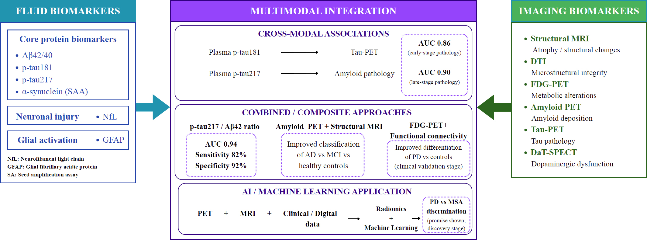
Integration of imaging and fluid biomarkers.
Okazawa et al. (2024) demonstrated that amyloid PET and DTI showed that deposition of amyloid peptides was significantly correlated with glymphatic clearance and structural brain alterations in patients with early AD, meeting the analytical validation requirements and warranting further replication [48]. The complementary use of these imaging techniques resulted in robust findings for the identification of neurodegeneration at early disease stages.
The combination of structural MRI and amyloid PET has been shown to further improve the accuracy of disease classification compared with single-modality imaging. A multi-centre study conducted by Bao et al. (2024) showed that the integration of amyloid PET with structural MRI measures significantly improved the differentiation between AD, MCI, and healthy individuals [49]. The use of structural MRI along with PET imaging provides complementary analyses that may support differential diagnosis in clinical settings, but analytical validation is still required for standardization. Further well-designed prospective studies are required to establish the findings observed.
The value of multi-modalities can also be extrapolated to PD, where differentiation may be established compared with healthy individuals. Li et al. (2025) showed that 18F-FDG-PET/MR can effectively reflect metabolic and functional alterations in PD patients. The combination of FDG-PET metabolic imaging and functional connectivity analysis yielded results that significantly improved differentiation between PD and control subjects, reaching the clinical validation stage [50]. The metabolic network connectivity may be represented as an imaging biomarker that can reflect neurodegeneration. Future multi-centre studies with a larger sample size are required to validate these findings.
Multimodal radiomics and machine learning have further shown diagnostic performance in distinguishing PD from other neurologic diseases. Sun et al. (2023) demonstrated that multimodal PET/MRI radiomics, combined with machine learning models, successfully distinguished PD from multiple system atrophy. This combination of PET/MRI radiomics-clinical machine learning models showed promising performance in discriminating PD from MSA, indicating scope for the development of future models for clinical use and positioning them at the discovery stage [51].
PET imaging is limited by accessibility and cost due to limited regional radiotracer availability. Additionally, repeated scanning may lead to cumulative radiation exposure. Quantification may be limited due to tau PET tracers binding to non-tau structures [46].
Imaging biomarkers provide structural context for the molecular and fluid biomarkers mentioned previously. CSF and plasma biomarkers provide biochemical insights into pathology that, when paired with structural MRI, functional connectivity, and PET imaging, localize that pathology anatomically and help track its spatial progression by visualizing processes such as tau spread, amyloid deposition, and dopaminergic decline. The consistent finding across multimodal studies is that combining structural, functional, and molecular imaging modalities improves diagnostic and prognostic accuracy compared with any single modality.
5. GENETIC AND MULTI-OMICS BIOMARKERS
5.1. Risk Genes and Pathogenic Mutations
Mutations in genes and susceptibility genes play important roles in the pathogenesis of neurodegenerative disorders. They influence protein aggregation, mitochondrial dysfunction, and neuroinflammation. Disease susceptibility is driven by monogenic mutations and polygenic risk variants.
In AD, mutations in the APP, PSEN1, and PSEN2 have been associated with familial autosomal-dominant early-onset disease that results in the production of amyloid peptides at an increased rate, which accumulate as plaques [52]. The APOEε4 allele is considered a strong risk factor for late-onset AD. Further, this contributes to impaired amyloid clearance and increased neuroinflammation [53].
In PD, gene mutations of SNCA lead to the accumulation of α-synuclein and the formation of Lewy bodies, which are representative hallmarks of PD pathology [54]. Gene mutations that are associated with mitochondrial function and impaired protein homeostasis, clearance, or α-synuclein aggregation, such as PARK2, PINK1, and DJ-1, collectively contribute to dopaminergic neuronal degeneration through oxidative stress and mitochondrial pathways [55]. Mutations in all the mentioned genes have reached the routine clinical use stage [53-55].
Genomic analyses in recent years have identified novel candidate genes that are associated with amyloid pathology, including CD180 and LY86, through a combination of transcriptomic and proteomic datasets. This suggests new molecular targets for exploration in neurodegenerative disorders [56].
5.2. Transcriptomics and microRNAs
New transcriptomic technologies allow a comprehensive analysis of changes in gene expression that are associated with neurodegenerative disorders. Wang et al. (2018) demonstrated that there are widespread alterations in genes that are associated with neuroinflammation, synaptic function, and mitochondrial metabolism [57].
RNAs such as microRNAs are being recognized as important regulators of gene expression in neurodegeneration at progressive rates. mRNA stability and translation are modulated by microRNAs, which thereby influence pathways that are linked to synaptic plasticity and neuronal availability. MicroRNA expression profiles being dysregulated have been detected in CSF and peripheral blood of patients with neurodegenerative disorders. The potential for microRNAs, therefore, warrants further investigation as minimally invasive biomarkers [58].
Transcriptomic studies in individual-level multi-omics have recently demonstrated that specific cell types, such as astrocytes and microglia, express distinct transcriptional and metabolic markers that are correlated with the progression of AD. The importance of specific cell-type gene regulation is therefore emphasized for consideration in neurodegeneration [59]. Transcriptomics and microRNAs are currently at the discovery stage [58, 59].
5.3. Epigenetic Signatures
Gene expression that are regulated by epigenetic mechanisms without changing the underlying DNA sequence are crucial to the pathogenesis of neurodegenerative disorders. DNA methylation and histone modifications are major epigenetic modifications in these disorders.
Significant epigenetic alterations in brain tissue and peripheral blood of patients have been expressed through studies of DNA methylation. Epigenetic alterations can affect the expression of genes that are involved in inflammation and neuronal availability. Further evaluation of these changes is required for assessment in neurodegeneration [60].
In PD, alterations in epigenetics have been associated with different methylation patterns in genes that are correlated with α-synuclein expression and mitochondrial function [61]. Increased α-synuclein expression and disease susceptibility have been correlated with hypomethylation of the SNCA promoter region. Currently, SNCA promoter hypomethylation is at the discovery stage. Overall, epigenetic alterations require further studies for evaluating their use as potential biomarkers for early disease detection and monitoring.
5.4. Proteomics and Metabolomics
Proteomics and metabolomics are crucial for providing insights into molecular alterations that reflect functional changes in neurodegenerative disorders.
Proteins that are associated with complement activation and neuroinflammation are correlated with amyloid accumulation and neurodegeneration. Wingo et al. (2019) demonstrated that circulating protein signatures are associated with AD pathology and subsequent cognitive decline. Proteins involved in mitochondrial activities or synaptic functions had increased abundance among subjects with stable cognition, with or without amyloid plaques or neurofibrillary tangles [62].
Emerging plasma proteomic panels, combined with machine learning, have been shown to differentiate dementia etiologies, including Alzheimer’s disease, frontotemporal lobar degeneration, and Lewy body disease, which is particularly relevant in clinical settings where these disorders present with overlapping symptoms [63].
Current alterations of metabolic pathways are associated with mitochondrial metabolism, lipid biosynthesis, amino acid metabolism, neurotransmitter metabolism, and hormonal metabolism in neurodegenerative disorders. Trushina et al. (2013) demonstrated that in MCI and AD, pathways related to mitochondrial function, lipid biosynthesis, amino acid metabolism, neurotransmitter metabolism, and hormone metabolism were altered canonically [64]. Circulating proteomic signatures and metabolomic panels are in the analytical validation stage and require further standardization. These changes can be reflected as indicators for early disease progression.
5.5. Systems Biology and Multi-Omics Integration
Systems biology and multi-omics datasets are combined to provide a comprehensive understanding of the mechanisms of neurodegenerative disorders. Integration of multi-omics enables the investigation of interactions among genes, transcripts, proteins, metabolites, and other substrates, and the identification of disease-specific molecular networks.
Integrated transcriptomic, proteomic, and neuropathological analyses have identified molecular signatures associated with amyloid pathology, neuroinflammation, microglial metabolism, and synaptic dysfunction in Alzheimer’s disease, positioning microglial biology as a candidate mechanistic driver and biomarker source at the discovery stage.
Multi-omics studies similarly integrated genetic and proteomic datasets, leading to the identification of biomarkers that are shared across multiple neurodegenerative disorders [62]. This suggests that altered molecular pathways, such as mitochondrial metabolism, immune activation, synaptic biosynthesis, astrocyte metabolism, and biosynthesis, may overlap.
Multi-omics datasets are increasingly being used by recent approaches such as machine learning and artificial intelligence for the identification of biomarker patterns, which can be used to detect early disease onset and enable progression suspension. Integrated approaches towards precision medicine and personalized therapeutic strategies in neurodegenerative disorders can be crucial for progression.
A comparative summary of omics-based biomarkers and their clinical readiness is presented in Table 3.
Despite promising outcomes, multi-omics integration is limited by inconsistent data standardization across platforms and substantial computational demands. These models are generally trained on small cohorts, raising overfitting concerns. There are limited panels that have undergone prospective validation in independent populations.
| Omics Category | Representative Biomarker(s) | Disease Context | Clinical Utility | Validation Stage | Major Limitations | Key References |
|---|---|---|---|---|---|---|
| Genomics (monogenic) | APP, PSEN1, PSEN2 mutations | Early-onset familial AD | Definitive genetic diagnosis; family risk counseling | Routine Clinical Use | Explains only a minority of AD cases (familial, early-onset); not informative for sporadic late-onset disease | [52, 53] |
| Genomics (monogenic) | SNCA, PARK2, PINK1, DJ-1 mutations | Familial PD | Definitive genetic diagnosis; family risk counseling | Routine Clinical Use | Explains a minority of PD cases; variable penetrance | [54, 55] |
| Genomics (risk allele) | APOE ε4 | AD | Risk stratification; trial enrichment | Routine Clinical Use | Risk factor, not diagnostic; ethical/psychological implications of disclosure | [52] |
| Genomics (polygenic) | GWAS-derived polygenic risk scores | AD, PD | Research risk stratification | Analytical Validation | Not clinically actionable at individual level; population-specific validity | [57] |
| Epigenomics | cfDNA methylation | ALS | Investigational diagnostic/prognostic marker | Discovery | Single-cohort data; Further studies are required | [24] |
| Epigenomics | CpG modifiers of disease onset | ALS | Investigational marker of onset timing | Discovery | No independent replication reported | [25] |
| Epigenomics | DNA methylation, SIRT/BDNF signatures | Dementia, PD | Investigational mechanistic marker | Discovery | Exploratory; Further efficacy studies are required | [26] |
| Epigenomics | SNCA promoter hypomethylation | PD | Investigational mechanistic marker | Discovery | Single recent mechanistic study; no clinical assay | [61] |
| Transcriptomics | Circulating miRNAs | AD, PD, ALS | Investigational diagnostic panel component | Analytical Validation | Expression profiles identified but no standardized clinical assay; cross-study inconsistency | [58] |
| Transcriptomics | Single-cell/hippocampal transcriptomic signatures | AD | Research tool for mechanistic subtyping | Discovery | Not yet framed for clinical translation | [59] |
| Proteomics | Circulating proteomic signatures | AD, PD, ALS, FTD (cross-disease) | Investigational cross-disease differentiation panel | Analytical Validation | Recent, largely single-cohort; independent replication pending | [62, 63] |
| Proteomics | Large-scale brain proteomic datasets | AD | Research resource for target/biomarker discovery | Discovery | Post-mortem tissue-based; not a fluid-accessible clinical marker | [75, 77] |
| Metabolomics | Plasma/CSF metabolomic panels | AD, PD | Investigational marker of mitochondrial/oxidative dysfunction | Analytical Validation | Pathway-level consistency only; no validated diagnostic cutoff | [64] |
| Multi-omics integration | Combined genomic-transcriptomic-proteomic-metabolomic panels | AD, PD (cross-disease) | Aspirational precision-subtyping tool | Discovery | No integrated panel has entered clinical validation; computational/standardization burden is substantial | [56] |
| Extracellular vesicle/peripheral omics | Exosomal, salivary, urinary multi-analyte panels | AD, PD | Investigational non-invasive panel | Discovery | Collection/standardization challenges; limited validation cohorts across all peripheral sources | [35-38] |
Multi-omics and genetic biomarkers add additional layers of mechanistic and risk stratification. While CSF, plasma, and imaging biomarkers provide comprehensive insights into the consequences of neurodegeneration, genetic and multi-omics data can offer insight into individual susceptibility, disease subtype, and the molecular drivers of pathology. However, the clinical readiness of these omics categories varies considerably, while proteomics and select genomic markers (APOE, SNCA) are approaching clinical-translational use, transcriptomics, epigenomics, and multi-omics integration approaches remain largely at an early emerging stage. Multi-omics provide value through precision stratification by identifying molecular subtypes within clinically similar patient populations that may respond differently to therapeutic interventions, rather than by replacing fluid and imaging biomarkers.
6. DIGITAL AND NEUROPHYSIOLOGICAL BIOMARKERS
The detection and monitoring of neurodegenerative disorders have been done using digital and neurophysiological biomarkers. Digital biomarkers, unlike conventional molecular biomarkers that rely on sampling or neuroimaging, can capture physiological and behavioural data using electrophysiological recordings and wearable sensors. Continuous monitoring of neural function and motor behaviour may be possible through these tools in analogous settings. Advancements in sensors and machine learning have recently significantly enhanced the ability to extract patterns relevant to diseases from complex datasets. Digital and neurophysiological biomarkers have consequently been used increasingly for their potential use in early diagnosis and disease monitoring of disorders such as AD and PD.
6.1. EEG and Electrophysiological Markers
EEG is a method for recording the brain's spontaneous electrical activity. It is a non-invasive method for measuring neuronal activity in the brain and has been widely explored as a biomarker in neurodegenerative disorders. In AD and PD, alterations in EEG spectral power and network connectivity have been observed. EEG recordings in AD frequently demonstrate abnormal activity and network dysfunction, is evidenced by increased delta and theta power and reduced alpha and beta activity. This is associated with synaptic dysfunction and neuronal loss [65].
In PD, electrophysiological biomarkers are being concurrently investigated. Decline in motor function has been linked to abnormalities in beta oscillations within cortico-basal ganglia circuits. Neumann et al. (2016) demonstrated that increased subthalamic 8- 35 Hz band power was strongly linked to clinical signs in PD patients. Low-beta frequency bands show a significant correlation with levodopa-induced reduction compared with high-frequency bands, suggesting variable functional roles for individual frequency ranges. Subthalamic oscillatory activity correlates with motor impairment, indicating that electrophysiological markers may provide informative insights into motor decline in neurodegenerative disorders [66]. EEG and electrophysiological markers are at the discovery stage.
High-density EEG and source localisation techniques have recently enabled the identification of disease-specific network disruptions by improving the resolution of electrophysiological analyses. Electrophysiological biomarkers, especially EEG-based, may complement imaging and molecular biomarkers for the detection and monitoring of diseases at early stages.
6.2. Wearable Technologies and Remote Monitoring
Monitoring of physiological and behavioral parameters has been enabled by wearable technologies in patients with neurodegenerative disorders. Detailed information on movement patterns and sleep behavior can be captured through devices equipped with accelerometers and physiological sensors. Motor symptoms in PD, such as tremor and bradykinesia, can be evaluated using wearable sensors. Del Din et al. (2019) demonstrated that motor activity obtained from devices worn on wrists was able to detect subtle motor impairments and predict disease progression in PD patients [67].
AD and MCI can also be monitored through remote monitoring technologies. Sleep disturbances and daily activity patterns being monitored continuously have been shown to correlate with cognitive decline and neurodegenerative pathology. Clinical symptoms of dementia preceding diagnosis can be detected through passive digital monitoring that is integrated into homes and observes daily routines. These wearable technologies require further validation and are currently in the analytical validation stage.
Advantages of wearable technologies, including objective quantification of symptoms and longitudinal monitoring, may allow for a reduction in reliance on clinical visits. Digital biomarkers collectively may prove to have utility that is particularly valuable for clinical trials with large populations and longitudinal disease monitoring.
6.3. Speech, Gait, and Cognitive Digital Phenotyping
Behavioral and physiological data generated through interactions with digital devices can be collected for analysis, which is referred to as digital phenotyping. Speech patterns and gait characteristics can serve as indicators of disease progression for assessment in neurodegenerative disorders.
Recently, speech analysis has emerged as a potential biomarker for neurodegenerative disorders. Symptoms such as reduced fluency and impaired articulation are generally observed as characteristic features in patients with AD and PD. The detection of cognitive and motor impairments in the early stages of the disease states is possible through computational analyses of acoustic features and speech timings. Lexical and syntactic features from narrative speech can distinguish individuals with AD from healthy individuals [68]. Early detection and disease monitoring through potential biomarkers, such as speech-based phenotyping, should be further evaluated.
Digital biomarkers such as gait analysis are also important. Motor coordination and balance generally lead to the development of measurable gait abnormalities in neurodegenerative disorders. Wearable sensors and motion capture systems identified specific gait signatures associated with PD, including reduced stride length and impaired postural stability [69]. Digital biomarkers that detect speech patterns and cognitive phenotyping require further real-world validation and are therefore at the analytical validation stage.
Cognitive tests based on smartphones have been shown to have promising results in the detection of early cognitive decline. Assessment of reaction time and memory performance, along with other cognitive assessments, may serve as objective indicators of cognitive impairment, enabling early detection of neurodegenerative disorders.
6.4. Artificial Intelligence in Biomarker Discovery
Artificial intelligence and machine learning approaches have recently emerged for the identification of complex biomarker patterns in neurodegenerative disorders. These approaches may be capable of combining multimodal datasets like neuroimaging and electrophysiological records for the identification of predictive signatures of neurodegeneration and disease progression.
Early AD pathology can be detected with high accuracy by analyzing neuroimaging data using deep learning algorithms. Neural networks that were trained on brain imaging datasets were capable of identifying individuals with preclinical AD multiple years before the onset of symptoms [70]. The analysis of motor activity decline in patients with PD for the assessment of disease severity can be performed through wearable sensors with machine learning models applied to them.
Biomarker discovery through artificial intelligence also enables the identification of previously unrecognized relationships between physiological signals and disease mechanisms. By analyzing data obtained through digital health technologies, machine learning models may aid in the discovery of subtle patterns that were undetectable through models based on conventional statistical approaches. Artificial intelligence and deep learning algorithms require further validation in realistic scenarios and are therefore classified as being at the analytical validation stage.
As access to data and computational power increases, artificial intelligence is expected to play a crucial role in the development of biomarkers for neurodegenerative disorders in the near future, allowing early-stage diagnoses of these diseases.
Data privacy concerns and regulatory pathways raise concerns for digital biomarkers. Interpretability may be subjective in artificial intelligence-based algorithms. Additionally, device heterogeneity limits cross-study comparability, and variable patient compliance introduces missing data.
Digital and neurophysiological biomarkers provide a niche role within the previously proposed multimodal framework. Fluid, imaging, and omics biomarkers capture a single time-point snapshot of underlying pathology, often obtained through invasive or resource-intensive procedures. Digital biomarkers obtained from EEG, wearable sensors, speech, and gait enable continuous, valid monitoring of motor, cognitive, and behavioral function in real-world settings. This complementary insight makes digital biomarkers well suited for longitudinal disease monitoring, remote clinical trial endpoints, and early detection of subtle functional decline between clinical visits that molecular or imaging biomarkers may not capture. Analysis is provided through artificial intelligence to combine multimodal datasets for detecting signatures of neurodegeneration that can be difficult to detect through conventional statistical approaches.
7. CLINICAL TRANSLATION AND IMPLEMENTATION
Translation of biomarkers from research to clinical practice is crucial for improving diagnosis and monitoring disease progression for neurodegenerative disorders. Pathological changes that are associated with diseases such as AD, PD, ALS, and MCI can be detected at early stages through molecular, imaging, digital, and multimodal approaches, significantly improving their prognosis detection. Challenges such as robust validation and regulatory approval need to be addressed for their implementation at the clinical level. Biomarker selection pathways for common clinical presentations, spanning cognitive, motor, and behavioral phenotypes, are summarized in Fig. (3).
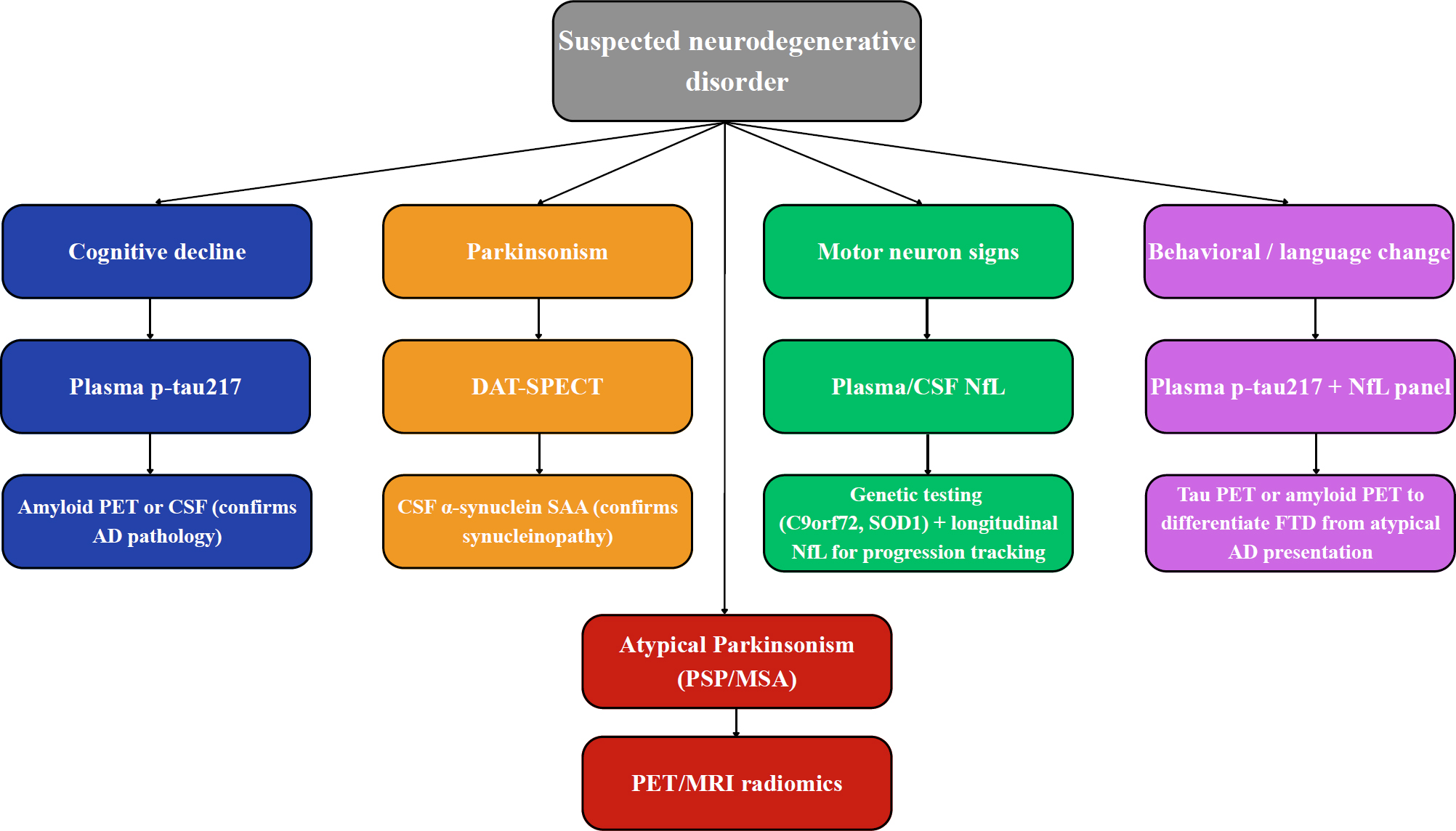
Biomarker selection pathways by clinical presentation.
7.1. Diagnostic and Prognostic Applications
Neurobiomarkers have gained increasing importance for their proficiency in diagnostic accuracy and specificity across neurodegenerative disorders, particularly at prodromal stages. Conventionally, diagnosis substantially relies on clinical symptoms, which appear at later stages of the disease when significant neurodegeneration has occurred. Duly, diagnostic approaches can overcome these challenges through the detection of these underlying pathologies at the preclinical or prodromal stages of disease.
As previously established, blood-based biomarkers can accurately identify pathological hallmarks of neurodegeneration. Plasma phosphorylated tau (p-tau217) produced high diagnostic accuracy for the identification of amyloid and tau pathology that directly correlated with AD. p-tau217 was demonstrated to be the highest predictor of cognitive decline in the group with Alzheimer’s clinical syndrome [71].
NfL can predict neuronal injury and disease progression across multiple neurodegenerative disorders such as MS, AD, FTD, ALS, atypical parkinsonian disorders, and traumatic brain injury. NfL should be complemented by other disease-specific biomarkers and brain imaging findings in clinical settings to realize its potential as a biomarker.
As discussed in Section 3.2, plasma p-tau217 has demonstrated the strongest cross-sectional diagnostic accuracy among current blood-based biomarkers for identifying AD pathology (AUC up to 0.96), while plasma p-tau181 has independently demonstrated both strong cross-sectional diagnostic accuracy and superior longitudinal prognostic value compared with T-tau, Aβ42/Aβ40, and NfL [9, 10]. This differential is clinically consequential: while NfL reliably signals that neurodegeneration is occurring, Section 3.2 shows it lacks the specificity needed to distinguish AD from other neurodegenerative diseases or to independently predict AD-specific risk, whereas p-tau species and CSF-based combinatorial algorithms (Section 3.1) can meaningfully reduce reliance on lumbar puncture and PET imaging for diagnostic confirmation [9, 10].
7.2. Biomarkers in Clinical Trials
Biomarkers play a crucial role in the design and execution of clinical trials in neurodegenerative disorders. Selection of participants, stratification, pharmacodynamic monitoring, and outcome measures are continuous use cases for biomarkers.
Biomarkers are often used to confirm the presence of amyloid or tau pathology for the selection of participants in AD-based clinical trials. This ensures that appropriate patient populations are selected for testing investigational therapies targeting specific pathological mechanisms. P-tau217 reduces the need for Aβ-PET imaging or CSF biomarkers in most individuals and minimizes the need for tau PET to identify individuals who will benefit from antiamyloid immunotherapies, improving trial efficiency and reducing costs [71].
NfL has been widely used as an indicator of neuroaxonal damage and as an endpoint for treatment in ALS-based clinical trials. Correspondingly, amyloid PET and tau PET are increasingly used as surrogate endpoints in trials evaluating therapies for disease modification before measurable clinical improvement occurs [45, 72].
Many clinical trials require biomarker evidence, such as Aβ pathology, for inclusion in AD trials. However, there may be interindividual variations due to different brain pathologies; therefore, it is important to determine the cause of symptoms for each individual. Subgroups that are presumably outlined by certain biomarkers may respond more to therapies.
Longitudinal studies focusing on these biomarkers that span from pre-symptomatic or prodromal stages of the disease are crucial for understanding how pathologies may overlap and manifest observationally, as well as their correlation with clinical symptoms and their differences.
7.3. Standardization and Regulatory Pathways
The standardization and validation of these biomarkers are necessary for their implementation in clinical practice. Changes or differences in laboratory techniques, sample collection methods, assay platforms, and imaging protocols may result in substantial variability across multiple studies. Inconsistencies in biomarker measurements can be produced due to the aforementioned variations, which may limit the reproducibility and clinical utility of these biomarker measurements.
International regulatory agencies such as the U.S. Food and Drug Administration and the European Medicines Agency have developed biomarker validation frameworks to assess the analytical validity and clinical validity of these biomarkers before they can be used in drug development or decision-making.
The importance of analytical validation in biomarker assays requires emphasis. Partial parallelism was generally inconsistent in its observation and reporting for most biomarker assays for neurodegeneration. Generalizability is limited due to the narrow range of dilution and inconsistent methodologies. Dilution protocols and adherence to attested analytical validation guidelines are of significant importance [73].
Cross-cohort validation of a plasma p-tau217 assay has been described as showing consistent identification of amyloid pathology across independent community-based cohorts, which is supportive of clinical implementation [74]. Standardization efforts, as previously demonstrated, need to be integrated into the validation of these biomarkers used for aid clinical diagnosis.
7.4. Cost-Effectiveness and Global Accessibility
Significant progress has been made in biomarker research. Despite this, the widespread implementation of biomarker technologies remains hindered by cost and global accessibility. Specialized equipment and trained personnel are required for techniques like PET imaging and Multimodal imaging, which increases their difficulty for implementation in healthcare settings.
Alternatives such as blood-based biomarkers are promising due to their affordability, scalability, minimal invasiveness, and diagnostic accuracy. Large diagnostic studies have shown that combined plasma Aβ ratio and phosphorylated tau assays can accurately detect Alzheimer’s disease pathology in both primary care and specialist settings, supporting their scalability outside dedicated memory clinics.
Recent developments have also highlighted the importance of blood-based biomarker tests for the transformation of clinical practice. p-tau217 has demonstrated high diagnostic accuracy for distinguishing Alzheimer’s disease from other neurodegenerative disorders in multicenter cohort studies [10]. This highlights important underlying frameworks that may aid with quality of life and patient-centred financial outcomes.
Digital biomarkers derived from wearable devices and remote monitoring technologies may improve accessibility through continuous monitoring of motor and cognitive decline outside clinical settings. Wearable sensors have demonstrated the ability to detect changes in gait and motor activity in patients with PD, providing valuable insights for disease monitoring and treatment [67]. Diagnostics based on biomarkers, including these technologies, have the potential to improve disease monitoring in healthcare settings with limited resources.
8. CHALLENGES AND FUTURE DIRECTIONS
Several scientific and practical challenges continue to limit the widespread clinical implementation of biomarkers in neurodegenerative disorders despite significant advancements in biomarker research. Heterogeneous clinical manifestations, variable rates of progression, and complex pathological mechanisms plague diseases such as AD, PD, ALS, and FTD. These factors, in combination with reproducibility and validity of biomarker measures and their translation into clinical practice, produce significant challenges. These challenges require addressing through large longitudinal studies and integration of technologies such as artificial intelligence and multi-omics analysis.
8.1. Biological Heterogeneity
Biological heterogeneity is a significant obstacle to the development of biomarkers for neurodegenerative disorders. Genetic susceptibility, complex neuropathological mechanisms, clinical presentations, and disease progression are confounding factors that need consideration in patients diagnosed with the same neurodegenerative disorder. The generalizability of candidate biomarkers may be limited by variability found in small discovery cohorts.
Large-scale multi-omics investigations have recently demonstrated that multiple molecular subtypes exist within neurodegenerative disorders, each characterized by distinct biological pathways. Neuroinflammation and synaptic dysfunction have been described as molecular signatures that are both disease-specific and shared across several neurodegenerative disorders, as suggested by cross-disease proteomic analyses. These findings suggest that underlying biological processes may develop seemingly similar clinical patterns of disorders.
Heterogeneity is evident, particularly in PD, where genetic mutations and neuropathological differences produce diverse phenotypes that may vary in motor symptoms, non-motor symptoms, cognitive impairment, and other clinical manifestations [75]. Neuropathological studies of Alzheimer’s disease have described distinct subtypes based on the regional distribution of tau pathology, differing in age at onset, genotype, and clinical trajectory [76].
Important challenges in biomarker development, such as heterogeneity, can be problematic, as diagnostic biomarkers must accurately detect disease pathology across diverse patient populations. Molecular subtypes are increasingly used for the classification of patients for stratification approaches, rather than relying solely on traditional clinical diagnoses. Integration of genomic, proteomic, metabolic, and imaging biomarkers may enable precise characterisation of disease subgroups and improve biomarker performance.
8.2. Validation and Reproducibility
The limited reproducibility of many candidate biomarkers may cause critical barriers to clinical translation. Numerous biomarkers demonstrate diagnostic accuracy in preliminary studies but may fail to replicate this when evaluated in independent cohorts or in practical clinical populations. Study design, sample collection, protocol adherence, and analytical methods may significantly influence biomarker measurements and contribute to inconsistent findings.
Factors such as standard operating procedures, sampling time points and methodologies, sample handling, and storage conditions have been shown to affect the stability and quantification of fluid biomarkers such as p-tau and NfL. Multicenter investigations have therefore emphasized the importance of adherence to standardised sample-collection protocols and randomised sample analysis to improve reproducibility across laboratories.
Computational approaches may also cause reproducibility challenges when used for biomarker discovery. Machine learning models derived from neuroimaging or multi-omics datasets using a single dataset are not replicated using other datasets. Dataset integration can result in a decrease in classification accuracy but also enhance the reproducibility of potential biomarkers. The average lack of reproducibility is significantly reduced by dataset integration, which is particularly important [77].
Addressing these limitations, international initiatives have established large collaborative datasets designed to validate biomarkers across diverse populations. Integration of datasets like imaging data, clinical phenotypes and genotypes, molecular biomarkers, and variance can produce robust resources for evaluating candidate biomarkers and improving their reproducibility.
8.3. Ethical Considerations of Early Diagnosis
Early detection of neurodegenerative disorders during preclinical stages, often multiple years before the onset of clinical symptoms, was made possible by advancements in biomarker technology. Recruitment of individuals into clinical trials that are generally high-risk for the detection of diseases raises important ethical and social concerns that need to be addressed.
The conceptualization of multiple neurodegenerative disorders has been fundamentally changed due to diagnostic frameworks based on biomarkers. The National Institute of Aging and Alzheimer’s Association (NIA-AA) has proposed a research framework that introduced a biological construct to define AD based on biomarkers that are specific to hallmark AD proteinopathies, such as amyloid deposition and tau pathology, rather than purely clinical criteria [78]. Similarly, recent proposals for redefining synucleinopathies emphasize that the accumulation of α-synuclein pathology should be the focus for considering synuclein diseases, even if they meet the clinical definitions of PD or DLB.
Complex ethical questions may still be raised despite the improvement in diagnostic precision. Individuals who may be positive for specific biomarkers may remain asymptomatic for several years or may never develop the indicated clinical disease. Expressing the existence of preclinical disease risk may result in psychological distress or social stigmatisation.
The potential misuse of biomarker information in employment or insurance settings may also raise additional concerns. Ethical guidelines should emphasize the importance of informed consent and genetic counselling. The benefits of early diagnosis need to be weighed against potential psychological and societal consequences led by mental disturbances, which may pose a central challenge to the implementation of diagnosis based on biomarkers.
8.4. Toward Precision and Preventive Neurology
The development of strategies for precision and preventive approaches to neurodegenerative disorders is guided by future advances in biomarker research. Precision neurology integrates molecular, imaging, digital, and fluid biomarkers with clinical data to identify disease subtypes and predict progression for individual patients.
Genomics and proteomics across diverse populations have been integrated to generate extensive biomarker datasets through global collaboration. Large datasets based on proteomics have been designed to identify biomarkers and therapeutic targets across multiple neurodegenerative disorders [79]. Large-scale resources, therefore, provide opportunities to investigate disease mechanisms and further develop accurate diagnostic tools.
Artificial intelligence and machine learning approaches are expected to accelerate biomarker discovery by enabling the integration of complex multimodal datasets. Combining data from neuroimaging, fluid biomarkers, genomics, proteomics, and digital monitoring technologies may allow clinicians to detect neurodegenerative disorders earlier and identify patients at higher risk of disease progression.
Finally, multimodal biomarkers combined with precision medicine may shift clinical practice from reactive treatment based on symptoms of the associated neurodegenerative disorder to preventive approaches that may delay or prevent disease onset. Achieving this, however, may require efforts for validate biomarkers and ensure ethical implementation on an ongoing basis.
8.5. Limitations of This Review
This review has several limitations that should be considered when interpreting its conclusions. As a narrative rather than a systematic review, study selection was guided by relevance and translational significance rather than by a predefined, reproducible screening protocol, introducing potential for selection bias in which studies were included and emphasized. The search was restricted to English-language publications, meaning relevant findings published in other languages may have been omitted, particularly from research communities where biomarker studies are conducted but not always translated into English-language journals. Coverage across neurodegenerative disorders is also uneven: because Alzheimer's disease has generated a substantially larger body of biomarker literature than Parkinson's disease, amyotrophic lateral sclerosis, frontotemporal dementia, or Huntington's disease, AD-related biomarkers are more extensively represented and more thoroughly characterized throughout this review than those for other conditions, which may understate the relative maturity of biomarker research in less-studied disorders. Furthermore, this review does not include a formal meta-analysis or forest-plot synthesis of quantitative findings; where diagnostic performance metrics are reported, they are drawn directly from individual studies rather than pooled across the literature, and readers should be cautious in generalizing single-study estimates, particularly where cohort size, population characteristics, or assay platform differ. Finally, given the pace of publication in this field, coverage of the most recent literature, including a number of 2025 publications, may be incomplete due to the lag between publication and indexing, and biomarkers or findings published after the literature search was conducted are not reflected here.
CONCLUSION
Neurodegenerative disorders such as AD, PD, ALS, and FTD represent growing global health challenges through progressive neuronal loss, complex molecular mechanisms, varying age of onset, and substantial heterogeneity. Advancements in biomarker research have significantly improved the collective understanding of biological processes underlying these disorders. Upcoming biomarkers based in molecular, genetic, imaging, fluid, and digital domains have transformed the diagnostic landscape, enabling earlier detection of disease pathology and providing valuable insights into disease mechanisms.
Molecular biomarkers such as protein aggregates, inflammatory mediators, metabolic signatures, and genetic influences, when used alongside neuroimaging techniques and fluid assays, have broadened the range of tools available for detecting neurodegenerative pathology. Biomarkers based on blood and peripheral approaches, such as saliva and urine, are being increasingly recognized for being minimally invasive and for ease of access. Supplementary technologies, such as wearable devices and remote monitoring systems, can enable continuous assessment of motor, non-motor, cognitive, and behavioural changes associated with neurodegeneration.
Despite these significant advancements, challenges associated with detection based on biomarkers need to be addressed before translation into clinical practice. Biological heterogeneity across subgroups, variability in disease progression, reproducibility issues, and methodological differences across studies make it difficult to validate these biomarkers. Ethical considerations regarding the psychosocial response to the disclosure of early disease detection must be carefully addressed, as biomarkers are increasingly enabling the identification of diseases at preclinical stages. These novel biomarkers need validation against neuropathology first, rather than being used in clinical diagnosis with lower precision.
Future research should focus on the integration of multimodal datasets such as neuroimaging, genomics, proteomics, metabolomics, and digital phenotyping, which can enable greater precision in the characterisation of neurodegenerative disorders. The implementation of artificial intelligence and computational modelling may facilitate future analysis of complex data to identify and predict patterns in previously unrecognized signatures. Individualised diagnosis and treatments, therefore, may be enabled through these approaches.
In the future, emerging neurobiomarkers may show promising results that can shift the methodologies for clinical practice with respect to their accuracy in detection and classification of neurodegenerative disorders, which can affect therapeutic strategies and possibly improve outcomes. By and large, biomarker research requires further global collaborative studies and standardized validation frameworks to address growing concerns about these discoveries. The integration of molecular, imaging, fluid, and digital biomarkers may ultimately pave the way for precision neurology, translating into clinical practice by shifting away from reactive treatment of neurodegenerative disorders toward early intervention and preventive strategies aimed at preserving neurological function and improving patient outcomes.
AUTHORS’ CONTRIBUTIONS
The authors confirm their contribution to the paper as follows: M.: Contributed to study conception and design; A.H.M: Drafted the manuscript and performed analysis and interpretation of the literature. Both authors reviewed and approved the final version of the manuscript.
LIST OF ABBREVIATIONS
| AD | = Alzheimer’s Disease |
| PD | = Parkinson’s Disease |
| ALS | = Amyotrophic Lateral Sclerosis |
| MS | = Multiple Sclerosis |
| DLB | = Dementia with Lewy Bodies |
| FTD | = Frontotemporal Dementia |
| MCI | = Mild Cognitive Impairment |
| GFAP | = Glial Fibrillary Acidic Protein |
| NfL | = Neurofilament Light Chain |
| Aβ | = Amyloid-beta |
| Aβ42 | = Amyloid-beta 1-42 |
| Aβ40 | = Amyloid-beta 1-40 |
| APP | = Amyloid Precursor Protein |
| p-tau | = Phosphorylated Tau |
| p-tau181 | = Phosphorylated Tau 181 |
| p-tau217 | = Phosphorylated Tau 217 |
| t-tau | = Total Tau |
| TDP-43 | = TAR DNA-Binding Protein 43 |
| PMCA | = Protein Misfolding Cyclic Amplification |
| RT-QuIC | = Real-Time Quaking-Induced Conversion |
| MRI | = Magnetic Resonance Imaging |
| rs-fMRI | = Resting-State Functional Magnetic Resonance Imaging |
| PET | = Positron Emission Tomography |
| SPECT | = Single Photon Emission Computed Tomography |
| DAT | = Dopamine Transporter |
| DTI | = Diffusion Tensor Imaging |
| FDG | = Fluorodeoxyglucose |
| CSF | = Cerebrospinal Fluid |
| DNA | = Deoxyribonucleic Acid |
| cfDNA | = Cell-Free DNA |
| RNA | = Ribonucleic Acid |
| miRNA | = MicroRNA |
| IL | = Interleukin |
| MCP-1 | = Monocyte Chemoattractant Protein-1 |
| MIP-1β | = Macrophage Inflammatory Protein-1 Beta |
| CRP | = C-Reactive Protein |
| TNF-α | = Tumor Necrosis Factor Alpha |
| Simoa | = Single Molecule Array |
| PSP | = Progressive Supranuclear Palsy |
ACKNOWLEDGEMENTS
Declared none.